Thursday, December 21, 2017

End-stage

“Dr. Sutton, have you taken a look at our next patient yet?”

I looked up from my computer to see my CA-2 resident standing in the workroom doorway.  “I was just pulling her chart, Joel.  How’s she look?”

“You’re not gonna like it, sir.”

It was 11 pm and our general surgery colleagues had posted an ex lap on a 55-year-old woman with free intraperitoneal air.  I opened her chart and started scanning her notes as Joel watched from the doorway.  One entry in her problem list stood out: alcoholic cirrhosis.  “How bad is it?” I asked.

“Horrible,” he said.  “Pull up her labs.”  He entered the room, followed by Kaitlin, the CA-3 resident on call.  Joel waited while the screen loaded, then pointed to a series of sequential BMPs.  “See that?  Right there, from one draw to the next.  Her chemistries looked stably marginal, then in the last two hours they just fell off.  Her kidneys just gave up.  And she’s on a non-rebreather upstairs with a gradually increasing oxygen requirement all day.  She’s in florid hepatorenal and hepatopulmonary syndromes.”

“You know I hate it when you’re right, Joel,” I teased.  Joel was an exceptional resident – one of the brightest and hardest-working people I’ve ever met.  I’d rarely found a reason to doubt his instincts or thought processes, and on this night I agreed with him completely.  Still, it’s my job to teach, so I swiveled my chair to face him and started to elicit his thoughts.

“What do you think we should do, Dr. Pierce?” I asked.

Without missing a beat, Joel told me his plan.  “I think we should ask the surgeons if they really think this is going to help.  Her perioperative mortality risk is off the chart.  She’s an optimal candidate for comfort care.”

“And if they say, ‘Damn the torpedoes’?” I asked.

“She’s already lined up,” he said, referring to the patient’s pre-existing arterial line and venous access sites.  “Induce with etomidate and a prayer.  Albumin for volume, get a gas and switch to product if indicated.”

“Extubate?” I asked.

He thought for a moment before answering.  “Maybe see how she does.”

I shook my head and pointed to her serial ABG results.  “She’s got progressive respiratory decompensation and they’re gonna do a big abdominal whack.  I think we’d get called to tube her at some point tonight regardless of whether she had surgery.  Let’s just plan to leave her intubated.”

“Makes sense to me,” Joel agreed.

I turned back to the computer and paged the attending surgeon, who returned my page almost immediately.

“Hi, Matt, you guys ready to bring our ex lap patient down?”

“Hey, Raji, I wanted to talk to you about that.  Her labs look like the beginning of the end.  Have you guys had a ‘goals of care’ discussion with her and her family yet?” I inquired.

“Yes, we did,” he answered.  “She’s got hepatic encephalopathy, but we discussed everything at length with her mother.  Her mom wants to go forward.”

I sighed.  “OK, they’re setting up room 6.  We’ll go see her and bring her down.”

Joel, Kaitlin, and I gathered the equipment we’d need to transport the patient to the OR, then went to the ICU to meet her.  We arrived to find a cachectic-appearing woman with clear increased work of breathing.  Despite the non-rebreather facemask, her oxygen saturation was 93%.  Her mother was at the bedside, an anxious woman who appeared to be in her mid-70s.  Two other women, who appeared to be the patient’s sisters, were also present.  A surgical resident was in the room writing orders, and two nurses were preparing the patient for transport.

As I took in the whole scene, I made introductions.  “Hi, ma’am, I’m Dr. Sutton and these are Drs. Berthold and Pierce.  We’re the anesthesiologists working tonight.  We understand the general surgeons think you could benefit from an operation.”

The patient nodded, but her mother answered for her.  “Yes, they think she has a hole in her intestines.”

I stepped to the bedside.  “We want to talk to you about your health and our plans for your anesthesia before we get going, OK?”

For the next couple minutes, my residents and I gathered information and learned little that we didn’t already know.  Her cirrhosis had decompensated to the point of impairing her pulmonary and renal function.  Otherwise, the patient and her family denied any significant medical issues.  Kaitlin and I stood back while Joel explained the anesthesia plan.  He told the patient she’d be intubated and remain unconscious throughout the operation.  Then he said that we intended to leave her intubated postoperatively.  The patient and her family went quiet as they digested this new piece of information.  After a moment, her mother spoke.

“How long will Patty have the breathing tube?” she asked.

Joel took a breath.  “We’re not sure,” he replied.

I stepped forward to take the lead.  “Ma’am, we sincerely hope she can wean off the ventilator over a period of hours and have the breathing tube removed.  But you’ve seen how her breathing has worsened throughout the day.  As sick as she is, there’s a chance that she might not be able to come off the ventilator at all.”

Again, the room went quiet for several moments.  “So she might have a breathing tube for the rest of her life?” the mother asked.  “For years and years?”

I looked around the room.  The nurses had stopped what they were doing and the anesthesia and surgery residents were still.  One of the patient’s sisters had tears in her eyes.  And in the mother’s eyes was a look of expectation and confusion.  She doesn’t understand, I realized.  She doesn’t know her baby is dying.  A pause.  Then, another thought: We’re having the wrong conversation.

I looked around the room.  When I spoke, my voice was steady but gentle.  “May I please have a few moments alone with Ms. Foster and her mother?”

The others in the room looked at one another before moving toward the door.  As the last of them left the room, I quietly sat facing the patient.  My words were addressed to the patient but directed toward her mother.

“Ms. Foster,” I began, “I need to be very frank with you.”  Both women eyed me anxiously.  I took a deep breath before continuing.  “I don’t think you’ll be able to wean from the ventilator, and I don’t think you’ll need long-term mechanical ventilation.  You’re dying, ma’am.”

Silence.  The faces of the patient and her mother registered unrestrained shock.  I had dropped a bomb, its impact just beginning to be felt.

The mother mouthed something before finding her voice.  Her next words were shaky as she grasped for hope.  “She can still get better, can’t she?”

My heart ached for this mother.  She'd clung to the hope that her daughter would recover, would live for years yet, and I had just torn that hope from her hands.  “I’d love it if I’m wrong,” I said, “but I don’t think so.  Everything that’s going on right now – this is the end of end-stage liver disease.”

Shock and silence before a feeble protest.  “But she saw her doctor six months ago, and he said she had plenty of time to turn things around.”

I shook my head gently.  “I can't say what she looked like then,” I answered.  “I can only base my opinion on how she looks now.”

“How long do you think, Doctor?  Months?”

Again I shook my head, and pulled out the last remaining foundation on which this mother had built her hope.  “Days.”

The mother's face was a picture of devastation.  Silently, I allowed her a moment to process my words.  Eventually, though, I had to go on.  “Ma’am, I’m sorry to have to give you that news.  The truth is, I don’t think your daughter will live much longer with or without surgery.  And we don’t have to do the surgery if you don’t want to.  We can initiate comfort care.”

“The surgeons said she would die without the surgery,” the mother said softly.

Just as softly, I answered, “She’s dying either way.”  I watched as the mother continued to survey the devastation of her world.  “Ma’am, your daughter’s liver has failed.  Her blood ammonia levels are very high.  That has the effect of impairing her judgment.  She’s not mentally capable of making an informed decision or consenting to an operation.  She has no other power of attorney.  You’re the one who has to make the decision.”  More silence.  Then, the final bomb: “Ma’am, I believe if we take her to the OR tonight, you will be called upon to decide – some time in the next week – to pull the plug and let her go.”

The mother stared at the floor in disbelief.  After several moments, I stood.  My face and voice conveyed my sympathy.  As a parent, my greatest fear is that I might have to bury one of my own children.  “I’m sorry.  I’ll leave you alone to think about things without outside pressure.  Take all the time you need.”  I stepped out of the room and closed the door.

My residents approached me and I told them what had transpired.  Then we stood in silence for the next five minutes as the heartbroken mother decided what to do.  Finally, the door opened.

“Please do the surgery, and do the best you can for her.”

I nodded.  “We’ll give her our very best, ma’am.”

Patty Foster survived surgery that night.  Six days later, with her mother and sisters at her side, she was compassionately extubated and allowed to leave this world.

Years ago, I was an eager young premed student.  I volunteered and was chosen to shoulder the mantle of a physician.  For years I trained, learning the art and science of my profession.  I've blessed thousands of lives, ushering people through the perioperative period.  Not a day goes by that I don't thank God for His goodness in making me a tool in His hand.  But with that gratitude often goes a plea: God grant that I not outlive my children.

Tuesday, October 2, 2012

Clinical detachment

I had just laid down when my pager went off.

“Hey man, we have an ex lap going into OR 18,” said my junior resident.

I looked at my watch.  The time was 2:45 AM.  I was still trying to shake the cobwebs from my head when my junior added, “ACLS in progress.”

That got my attention.  “Room 18?” I confirmed.  “I’ll be right down.”

I hurried to the OR and grabbed a mask, looking through the window as I did.  Despite the flurry of activity surrounding the patient, I could see that the left side of his chest was cut wide open.  I could see his heart beating through the incision.

People were already engaged in resuscitation as I entered the OR.  I quickly surveyed the scene – patient status, monitors, lines, etc. – as I stepped to the head of the bed.

“What’s the story?” I asked my attending.

“This guy fell forty feet from a train,” he replied.  “Get an a-line started.”

I grabbed an a-line start kit and an ultrasound as I stepped around to the patient’s arm, anticipating difficulty finding a good radial pulse.  “He fell off a train? Strange place to be at two in the morning.”  I felt for a pulse, got nothing, and fired up the ultrasound.  In place of a pulsatile artery, I saw a shallow round vessel that was slightly less compressible than its neighbors.  After two attempts at cannulation, I was rewarded with sluggishly pulsing arterial blood.

By the time I secured the arterial line, the surgeons had already opened the abdomen and unleashed a torrent of blood.  I stepped to the head of the bed and began directing the patient’s resuscitation together with my attending.  The first ABG was not reassuring.  pH 6.75, PaCO2 over 70, base deficit approaching 30, iCa around 0.7...things were looking less than stellar for this patient.

Over the next several minutes, we continued resuscitating the patient with blood products, vasopressors, calcium, and other drugs, attempting to keep up with the rapid blood loss that continued on the other side of the drape.  I asked my attending what else had transpired prior to our operative management, and he told me that the patient had arrived in the ED with cardiac activity but without a palpable pulse.  He quickly proceeded to complete cardiac arrest, at which point a code was called.  The ED attending was ready to pronounce the patient, but the chief surgical resident on call performed an emergency thoracotomy, applied an aortic crossclamp, performed manual cardiac massage, and injected two doses of intracardiac epinephrine, at which point the patient recovered a perfusing rhythm and was rushed to the OR for surgical management.

“What do you see in there, John?” I asked the surgery resident.

“He’s got a huge liver laceration,” he answered.

“Is he making any clot for you?” I asked.

“Not really.”

So we continued with our management, administering packed red blood cells for oxygen-carrying capacity and plasma, platelets, and cryoprecipitate to promote clotting.  But the bleeding was too fast, and we seemed to be falling behind.

“Give him factor VII, 90 mcg per kilogram,” ordered the attending trauma surgeon.

Factor VII is a recombinant clotting factor.  It is reserved for cases of severe coagulopathy in which other attempts to control bleeding have failed.  This is for two reasons: it can promote dangerous amounts of clotting in many patients, and it costs about $1 per microgram.  The requested dose of 9 mg was therefore something to be given only when necessary.  We called the pharmacy and placed the order.

After a few minutes, the factor VII arrived and was given.  Still the patient continued to bleed.  Still the surgeons attempted to control the hemorrhage.  Still we persisted in our resuscitative attempts.  The trauma surgeon ordered 10 mg more factor VII, and this was given.

And then the trauma pager sounded.

“Major trauma alert,” read my junior as he took the pager from his hip.  “Gunshot wound to chest.”

“I know about that patient; he’s stable,” said the trauma surgeon, “but I should still go eyeball him.”  With that, he scrubbed out and left the OR.

For the next ten minutes, we continued our resuscitative efforts as the surgery resident packed the patient’s abdomen in an effort to tamponade the bleeding.  But when the attending surgeon returned, we still seemed not to have made much headway.  And in addition, the patient’s pulmonary status was worsening, probably from all the blood products he’d received.

“How’s it going, John?” the attending surgeon asked as he re-gowned.

“Um...not excellent, sir,” the surgical resident responded.  “He’s still bleeding pretty bad.”

“He’s not looking great up here, either,” I told him.  “We’ve got increasing airway pressures and decreasing sats.  I haven’t been able to suction anything out of his tube, either.”

Our resuscitative efforts continued in earnest as the attending surgeon and attending anesthesiologist conferred.  Ultimately, they decided that the best course of action would be to pack the patient’s abdomen and take him to the ICU for continued resuscitation and respiratory optimization on an ICU ventilator.

My attending and junior transported the patient to the ICU while I stayed behind in the OR to reconcile the chart and ensure that all blood products and controlled substances were documented appropriately.  It took me over an hour, and I eventually determined that the patient had received over 80 units of blood products in our attempt to save his life.

Unfortunately, there is no happy ending to this story.  The patient required more epinephrine upon arrival in the ICU and another code was called within minutes.  Within forty-five minutes of leaving the OR, it was all over.

The next day, I went to church.  I spent some time talking to a friend and told him about my patient.  By the time I finished, he was shaking his head.

“I don’t know how you guys do it.”

“Do what?”

“How you handle taking care of these sick patients without losing it?  I couldn’t handle it.”

I had to think about his question a bit before I could answer.  While I’ve been involved in the care of several patients who have died, none has affected me quite as much as the first one did.  After this most recent patient death, I had finished my charting, driven home, and gone to bed.  Was that callous?  Had I become insensitive to the sanctity of life, able to attend its passing with as little emotion as I would feel watching the “death” of character played by a television actor?  The patient who had just died was someone’s friend.  He was someone’s son.  Maybe he was someone’s brother.  Maybe their husband.  Maybe their father.

So how did I deal with my proximity to such tragedy?  I maintained clinical distance from the situation, viewing the patient as a case rather than as a man.  And even as I realized this with a wave of guilt, I knew that’s how it had to be. 

Maintaining clinical detachment from tragedy in the hospital is how I maintain the composure and professional objectivity to make decisions under pressure.  It’s how I keep my nerve when I know a poor decision could result in considerable harm or death to another person.  It’s how I reset after a difficult or complicated case so my next patient can receive my full attention and best care.  It’s how I fall asleep mere hours after watching the life leave another human body.  I can’t speak for my colleagues, but emotional detachment from tragic circumstances is the coping mechanism that helps me face serious illness and say, “I can handle this.”

And yet, in my quieter, more reflective moments I often feel the humanity of my work rush back to the forefront of my consciousness.  I find myself regretting my inability to cure all ills, and I pray that God will right the wrongs that defy the limits of human ability and of mortal comprehension.  It’s during such times that I remember the faces and the voices of those who have slipped beyond the veil of this life and into the next.  I pray that they have peace, and that their loved ones may find comfort even as they grieve.  I think of my own father and give thanks for the outstanding medical care that eased his pain at the end of his mortal journey, and I pray that I may have the wisdom, knowledge, and sound judgment to provide such excellent care even when faced with disease that exceeds my ability to heal.

During these reflective moments, the patients cease to be cases; they again become human beings, men and women whose time on Earth came to an end.  Inevitably, I reflect on the tenuousness of life and of the common threads that tie all of us together, the events and needs that we all experience.  Then I realize that each patient who dies is not just a patient; it is my brother.

And with that realization, I have wept for those I was unable to heal.

Tuesday, September 18, 2012

Do I look stupid to you?


Spontaneous rupture of membranes (aka SROM, aka water breaking) is a great reason for doctors to actively intervene in a pregnant patient’s care.  If the pregnancy is at term, it’s a good time to induce or augment labor in order to facilitate childbirth.  If it’s preterm, it’s a good time to attempt to stop or prevent labor, at least temporarily, to help prepare the mother and baby for delivery.  If, on the other hand, the water has not broken but the mother tells us it has (either because she really thinks her water broke or because she’s just tired of being pregnant), it’s important that we not promote labor and delivery, largely for the benefit of the child.  Either way, if a woman tells us her water has broken, it’s important for us to determine whether or not that’s really the case.

There are, of course, ways for the OBs to tell if a patient’s SROM is legit.  Typically, they perform three tests: speculum exam, ferning test, and nitrazine test.  The speculum exam involves using a speculum (what else?) to look for pooling of amniotic fluid the patient’s cervical vault.  The ferning test is done by putting a sample of the presumptive amniotic fluid on a microscope slide and looking for a characteristic crystallization pattern.  And the nitrazine test is done by exposing a sample of presumptive amniotic fluid to nitrazine and watching for a distinctive, characteristic color change.  If more than one of these tests are positive, it’s more likely that the patient did indeed experience SROM.

Despite modern medicine, however, some TOBPs think they can fake out their OBs.  "Hmm," they think, "I'll bet if I sit on a gurney and get the Chux pad under my butt wet, the doctors will think my water broke."  And what's the easiest way for them to get that pad wet?  Why, urinating on it, of course!

Which brings us to the patient.  She was a young woman, maybe 25 years old, who presented to the L&D deck one night while I was covering the night OB anesthesia service.  She arrived with a large water bottle in hand (really a 32 oz. mug full of water), which she was quite attached to.  She was complaining of regular, severe contractions, despite appearing to be quite comfortable.  The L&D nurses set her up in a triage room and took her vital signs, then she was seen by an OB resident.

The OB resident emerged from the patient's room several minutes later, shaking her head.  "That patient is gross," she said.

"What happened?" I asked.

"I was getting ready to do a spec exam, and I had my back turned to her, and she peed all over the Chux!" was the disgusted reply.

"Wait, what?!"

"She peed!  I had my back turned, and I was putting on gloves, and she got all excited and said, 'What was that?!  What was that?!  My water just broke!"

"OK..." I prompted.

"The Chux pad under her was wet, and her urine was really dilute from all the water she's drinking, but I could smell it and there wasn't much fluid in her vagina.  She peed on the Chux to try to make me think she SROMed."

"Wow..." I muttered, shaking my head.

"Anyway, I got what sample I could, and now I have to check nitrazine and ferning.  See you."

With that, the resident left and I returned to my reading.  I had all but forgotten about the incident when the OB resident returned and tapped my shoulder.

"OK, Matt, get this: you know how sperm makes the nitrazine test have a false positive?" she asked.

"You're asking me to cast my mind back to the dark days of med school, but yeah," I answered.

"Her nitrazine turned blue, but I could see swimmers on the slide.  So I went back in and asked her when the last time was that she had sex."

"OK..."

"And her husband said, 'Oh, it's been more than a week,' but she got really quiet and wouldn't look at me."

I looked over at the resident.  "You don't think..."

"Yes, I do think.  This lady is some piece of work.  I think I'm not the only one she's lying to this evening."

I shook my head in disbelief.  "I guess I'd rather be in your shoes than her husband's.  You think he'll put two and two together, and figure out why you asked when they had sex?"

"Maybe," she answered.

And there you have it.  Sometimes, the answers you get are a bit more than you bargained for.  And as irritating as it is when patients think their doctors are gullible fools, there are certainly worse things.

Tuesday, September 4, 2012

Ascending aortic dissection


No matter how much you know about medicine and anesthesiology, you’re going to come face-to-face with cases that challenge you.  Whether it’s the patient’s disease process, their overall health status, or considerations stemming from the surgical procedure itself, some cases are going to present a challenge.  And when those factors all combine with patient beliefs that prevent you from using some of your most important tools, you just set up for the case and know that you’re screwed six ways to Sunday.

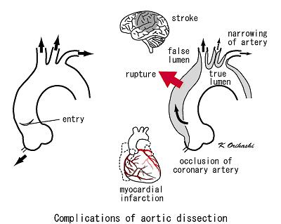
My patient was a 33-year-old black woman with Marfan syndrome.  She had suffered an ascending aortic aneurysm with subsequent dissection.  Her chart showed that she had presented to an emergency room two months prior for chest pain, but a full workup had been unrevealing.  At the time of her dissection, she had become neurologically compromised, and on the day of her surgery she was in the ICU intubated but requiring no sedation.  She had also become hemodynamically unstable and her kidneys had failed; she was on CRRT.  She had anasarca with her tongue so swollen that it protruded several centimeters from her mouth; it was obvious that if we dislodged her endotracheal tube she would asphyxiate as we were unlikely to be able to replace it.  Similarly, we could see that placement of her lines was going to be very complicated.

The surgical plan was, of course, for median sternotomy, cardiopulmonary bypass, and repair of her dissected aneurysm.  Bypass causes notable dilution of the patient's blood, and transfusion of red blood cells and other blood products is very common in such cases.  This patient, starting with her hemoglobin a very anemic 7.3 gm/dL, would certainly require transfusion.

Except that she was a Jehovah's Witness.

It's not much fun to walk into a case knowing that your patient will die if she doesn't have surgery, but that she'll probably die because of the surgery.  The only solace you can take is from the knowledge that no matter what you do, it's going to be very difficult for you to make her any worse.  Some consolation.

The procedure and anesthetic management had been discussed at length with the patient's family, and their wishes had been made extremely clear.  We documented everything imaginable, partly so we could honor the patient's and the family's wishes and partly so we would know what fluids we would be able to administer.  The patient could receive no red blood cells, platelets, or plasma, but she could receive albumin, recombinant factor VII, and Cell Saver.

My attending and I made the best of it and prepared for the procedure.  He handled transport since my mobility is still limited by my tibial fracture.  The anesthesia techs helped with positioning and monitoring the patient, and I scrubbed and gowned for the placement of lines.

This patient already had a triple-lumen central line in her left IJ and it was simple to swap it over a wire for one of the double-lumen lines preferred at this particular hospital.  Placement of a MAC in her right IJ was a bit more complicated due to her extreme edema.  With virtually no neck mobility and great difficulty in retracting her edematous folds of skin, a two-handed procedure became a four-handed one as my attending and I each retracted skin in opposite directions, he handled the ultrasound, and I drove the needle.  We ultimately prevailed, though, and the case was soon underway.

Transesophageal echocardiography is a very useful tool in assessing the anatomy and function of the heart and the great vessels.  This young lady's TEE was wretched.  It showed a functional heart pumping blood into a badly traumatized aorta, a massive communication evident whereby blood could move from the true lumen to the pseudolumen.  The pseudolumen had grown in size until it dwarfed the true lumen; I thought it miraculous that the patient still had peripheral pulses.  The surgeons and my attending, for whom cardiac surgery and cardiac anesthesia are daily aspects of life, seemed impressed by the extent of her dissection.

For several hours, the surgeons worked, opening the young woman's chest and repairing her aorta.  All along, there were indications of how sick this young woman truly was.  Her BIS was constantly low, starting in the mid-20s when we rolled into the room and actually going to 00 with a flat EEG on 0.3 MAC of isoflurane, indicating the probable extent of her neurological devastation.  We drew no unnecessary lab work, regarding every milliliter of her blood as a precious commodity that should not be wasted.  An ABG drawn shortly after initiation of bypass showed that she was severely anemic, with a hemoglobin concentration of about 6 gm/dL.  Instead of waiting until the end of the case to administer Cell Saver blood, we asked the perfusionist to start making whole and half units as soon as he was able.

And still the surgeons worked.  They were ultimately successful in repairing her aorta, and we all breathed sighs of relief as the patient tolerated the weaning of cardiopulmonary bypass with only two vasoactive infusions.  The sternotomy was closed and the attending surgeon scrubbed out to dictate the case as his assistant continued with skin closure.

But as he closed, the patient began to deteriorate.  Her pressures worsened, her heart rate accelerated, and her BIS, already tenuous, dropped.  Fearing what we would see, my attending and I looked at the TEE and found the diagnosis: cardiac tamponade.  The patient was bleeding into her pericardial sac, and the blood looked like it was already clotting.

We showed the junior surgeon what we had seen and he immediately began cutting the sutures holding the patient's chest closed.  The attending surgeon hastily scrubbed back into the case and within a minute the patient's sternotomy had been reopened.  My attending and I watched the surgeons scoop clotted blood from the patient's pericardium, knowing that every red blood cell lost to that clot was a cell she needed desperately.

 As the surgeons closed the patient's chest again, we very cautiously administered albumin to maintain a reasonable blood volume without hemodiluting the patient too much.  By the end of the case, the patient's total fluid input was just one liter of albumin and 300 ml of lactated Ringers.  And still her final hemoglobin was just 4.4 gm/dL.  My attending and I traded dire prophecies with one another after we dropped our patient off in the ICU.  Neither of us thought she would live out the night.

Nearly a week later, she remains alive.  But all is not well.  Her neurological exam remains remarkably poor, and each day further dims the hope that she will experience any sort of meaningful recovery.

To me, that is one of the most difficult parts of medicine: knowing you've done your best, knowing you've done as well as anyone anywhere could do, and still feeling that you've been beaten.  All you can do is say a prayer for your patient and hope that you can help the next one.

Monday, August 20, 2012

Ortho to the rescue!

So, I've given Ortho some grief on my blog, and I think it's time to give them their due.  That's largely because of my innate sense of fair play, and also because they recently saved me from serious injury.

I've never been much of a runner, but I decided to take it up in earnest about six weeks ago.  My decision was made partly to trim a little from my waistline, partly for cardiovascular fitness, partly for bragging rights ("my 5k time is better than yours"), and partly because I was on an out-of-town rotation and staying in an apartment complex with an air-conditioned workout center with treadmills.  I started fairly slow -- just 1.5 miles on each of my first few runs, building up over the course of a couple weeks to a bit past 2 miles.  Nothing out of the ordinary -- not even that long of a run.

My mistake was to run too frequently from the beginning.  My previous attempts to become a runner involved a MWF jogging schedule, adjusted as necessary based on my work schedule, but never with more than three runs per week.  While I was out of town, however, I had lots of free time and none of my normal distractions (family, video games, my movie collection, etc.) so I adopted a M-F running schedule with rest on Saturday and Sunday.

About two weeks into this schedule, I began to develop some left knee pain, particularly at the beginning of my run and after it was finished.  I foolishly rationalized that it was simply muscular soreness that would resolve with time in my new routine, so I continued my aggressive running schedule and used Motrin 800 mg tid to control my discomfort.  To an extent, it worked, and I was able to keep pushing myself more than I should have done.

My away rotation was four weeks long, and by the time it was over I realized something more than simple muscular soreness was at work.  I backed off to two runs per week, but those had become very painful, especially for the first half mile.  After each run, my wife was treated to my best Vanilla Ice impression as I collapsed into a chair ("Ice, ice, baby!").  My knee pain had quickly progressed from something associated with my runs to constant discomfort, hurting me even on my run-less days.  I was loathe to stop running, since I had come to enjoy it apart from my knee pain, but something was clearly wrong with my knee and I knew I needed to back off or risk serious injury.

At this point, I had a quandary: should I use my own medical knowledge to manage my injury conservatively, or should I involve an orthopedist?  It's not always easy for doctors to get time away from their clinical responsibilities, especially if they're residents.  Fortunately, one of the perks of medicine is the ability to informally ask for medical advice from colleagues in many specialties, and I approached an orthopedist in the OR two weeks ago to ask his recommendation.  He replied that he'd be happy to examine my knee in between cases, and when he did he became a bit concerned.

"You have a clear medial collateral ligament strain," he told me, "but you've also got pain too anterior to be explained by that.  I'm worried you may have a small medial meniscal tear."

The orthopedist (I'll call him Dr. Thompson) ordered x-rays and an MRI to further evaluate my injury, asking that I call him when the scans were done so he could check them.  I was able to get the x-rays done the same day, but MRIs must be scheduled and it took a week before I was able to have that study completed.

On the appointed day, I went downstairs for my MRI scan.  It was completed in about 30 minutes, and afterward the technician returned me to the waiting room while the radiologist checked the images to confirm their adequacy before I left the MRI clinic.  Five minutes later, the technician came back and informed me that the radiologist was on the phone and wanted to speak to me.

"Hi, this is Dr. Wong," he said.

"Hi, I'm Matt, one of the anesthesia residents," I replied.

"Matt, I'm looking at your MRI right now and you have a grade 4 stress fracture of your medial tibial plateau.  I want to get you on crutches.  This fracture looks pretty impressive."

I had a sinking feeling, knowing how crutches would complicate my job, which includes pushing patients around the OR suite and spending a lot of time on my feet.  But two things the radiologist said caught my attention.  First, he said the fracture was grade 4.  I was unfamiliar with the criteria for grading stress fractures by MRI, but lots of things in medicine are graded on a scale of 0 to 4 with 4 being the worst.  Second, he said the fracture looked "impressive."  That's generally doctor-speak for, "damn, that looks bad!"

Just to be sure, though, I asked him what a grade 4 fracture was.  He said it meant you could actually see a fracture line on the MRI instead of just bone marrow inflammation and edema.

So off to the radiologist I went.  He showed me my MRI (the picture at the top of this post is a screen capture from my MRI, and all the white crap in my tibial plateau is edema and angry inflammatory cells that shouldn't be there) and wrote me a prescription for crutches, which I could pick up from Physical Therapy.

A few minutes after I picked up my crutches, my cell phone rang.  It was Dr. Thompson, who had already looked up my MRI.  He had taken the time to look up my home number from the hospital computer system, then had my wife give him my cell so he could touch base with me.

"Matt, this stress fracture isn't pretty," he said.  "You need crutches."

"Just picked them up, sir.  Dr. Wong from radiology said the fracture looked ugly."

"Yeah, it's impressive.  In addition to crutches, I want you in a full-length knee brace.  It should be unlocked because I want you to be able to move your knee, but you need to wear the brace at all times.  And you can toe-touch, but no weight-bearing at all on that leg."

That gave me another indication of just how much damage I had done to my knee.  Total non-weight-bearing?  For an active resident who has to walk around the OR suite and the hospital all day long?  That's serious business.

I asked about the other MRI findings and Dr. Thompson told me that the MRI confirmed the MCL strain he'd diagnosed, but my meniscus looked good.  He told me where to go for the brace and said he'd follow up with me on his next operating day.

And that's pretty much where we are now.  I'm hobbling around on crutches because I broke my leg by advancing my running schedule too aggressively.  But for now, I seem to have avoided more serious injury (like the fracture extending all the way through my tibial epiphysis).  I know the extent of my injury and I now know exactly why my knee was hurting me so much, which knowledge is empowering since it allows me to undertake the appropriate management to promote healing.  And it's all because a good, attentive orthopedist was willing to increase his own workload by examining me between his cases, then take an active role in my follow-up and intervention.  And a good radiologist was willing to stick his neck out, too, instead of just dictating his findings and closing the MRI.

So here's a big "thank you" to Drs. Thompson and Wong (whose names have been changed, as always) for their excellent work in diagnosing and treating my fracture.  I'm deeply grateful for their willingness to go beyond the call of duty to follow up with me and ensure that I received the information and equipment required to prevent further injury.

And I learned a lesson from all this: when you decide to take up running, it is important, as with so many things in life, to pace yourself.

Monday, August 13, 2012

How to become a doctor

Most laypeople don't understand the medical education process.  When I was a med student, lots of folks asked me what my specialty was; now that I'm a resident, people sometimes ask when I graduate and become a doctor.  I've spent a lot of time clearing up confusion, often with varying degrees of success.

The pathway into medicine starts in high school or earlier.  Anyone who thinks he or she might like to be a doctor needs to perform well enough in high school to secure college admission, preferrably at a reputable school with a good track record for sending graduates to professional (MD/DO, DMD/DDS, PhD, etc.) programs.  Smart high school students will take a tough courseload with multiple AP and math or science classes.  Over my final two years of high school, for example, I took seven AP courses in subjects ranging from US history to calculus and from English literature to physics.

Then comes college.  Some universities have a dedicated premed major, others don't; but prospective doctors may major in any field they desire, so long as they remember that medical schools have specific requirements for applicants.  Med schools typically require several science and math courses, as well as other courses like literature and philosophy to ensure a well-rounded application.  Premed students also need to consider the content of the Medical College Admissions Test (MCAT), the standardized exam required of all med school applicants.  The MCAT tests physics, biology, and organic and inorganic chemistry, as well as verbal reasoning and writing abilities.  So premed students can major in French or journalism if they'd like, but they'll need to fill up their elective slots with coursework that will both prepare them for the MCAT and satisfy med schools' admission requirements.

As strenuous as these requirements may seem, there are still far more outstanding prospective physicians than there are med school spots, so schools also consider non-academic pursuits as a way for applicants to set themselves apart.  Schools are interested in applicants who are involved in the community, who are engaged in service opportunities, and who participate in extracurricular activities like music and sports in addition to maintaining a full courseload with excellent grades.  They also want applicants who have shown some dedication to the medical field, whether it's going on an overseas humanitarian mission or simply shadowing a local physician after school.  Healthcare-related employment is helpful.  And having published research on your CV is a big plus.

The next step to becoming a doctor is to apply to med school.  Some universities (like the one where I did my undergrad work) have a premed committee that helps guide premed students through this process, advising them about application requirements, counseling them about their chances at different schools, and organizing their letters of recommendation and other supporting documents.  A year before the applicant wants to start med school, the student fills out an online application through the American Medical College Application Service (AMCAS), a service operated by the Association of American Medical Colleges (AAMC).  Nearly all med schools in the nation accept the AMCAS application (the University of Texas system has its own application service), and the applicant simply checks boxes for the schools to which he or she is applying.  There's a fee for the first school checked, then a flat fee for every school afterward.  The AAMC notifies the university premed office of the schools to which the application has been submitted, and the committee forwards the applicant's packet to those schools.

Shortly after submitting the primary application, a prospective med student will begin receiving letters from the schools to which he/she applied.  These letters contain instructions for completing each school's secondary application, which contains individualized questions not covered by the primary AMCAS application.  And each secondary application must be submitted together with an additional application fee; when I applied to med school in 2004, the fees were anywhere between $50 and $100 for each school.

And then comes the waiting.  Each med school reviews the primary and secondary application, together with the supporting documents.  They weed out the applicants in whom they have no further interest; these students receive a letter thanking them for applying and wishing them better luck elsewhere.  The remaining students also receive letters inviting them to visit the med school for an interview.  It is up to the applicants to schedule their interview date, get themselves to the school, make it to the appropriate location on their interview day, and effectively sell themselves to the school.  This can get quite expensive, and applicants often apply to several schools in a given area so they can (hopefully) knock out multiple interviews in a single trip.  The interviews themselves vary in terms of their content and focus; but they all share the similarity of being a final chance for each applicant to set him or herself apart from the thousands of hopefuls who will not be accepted.

After the interviews come more waiting.  Starting in October, med schools start sending out letters telling each interviewee their status: in, out, or wait-listed.  Some applicants receive one admission out of all the schools they applied to; some receive multiple admissions; and most receive none.  Applicants are allowed to accept multiple admissions, but there is a date in the spring by which they must relinquish all but one admission so schools can determine the fates of the applicants who were placed on the wait list.

Med school starts a few months later, generally in the mid- or late summer.  A traditional med school curriculum is four years long and is divided between two years of classroom learning and two years of clinical work.  During the classroom years, students spend several hours each day in lectures and small workgroups, then spend a large portion of their out-of-school time studying.  There are frequent exams, which are uniformly rigorous.  Med students also frequently find themselves in the hospital during the evenings, learning the basics of hands-on patient care (how to take a pulse, how to check blood pressure, how to take a medical history, how to perform a physical exam, etc.).

At the end of the second year of their training, med students are required to take Step 1 of the United States Medical Licensing Examination (USMLE, aka "the boards").  It's a day-long standardized exam involving about 350 questions, and it must be passed before the student is permitted to assume clinical duties as a third year med student.  The exam has long-term implications, too: just as universities consider SAT/ACT scores and med schools look at MCAT scores, residency programs use board scores to determine which prospective residents interest them.  Since it is such an important exam, many schools provide their students several weeks without coursework so they can focus solely on studying for Step 1.

The third year of med school is often the hardest.  Med students spend their time in the hospital, passing several weeks at a time on each rotation before moving on.  My med school required third-year rotations in internal medicine, general surgery, pediatrics, family medicine, obstetrics/gynecology, psychiatry, and neurology.  Students receive grades for their performance on rotations; these are generally subjective grades, but some services also have a written or oral examination that factors into the grade.  The grades are important, as residency programs see them and use them to decide between applicants.

After they've finished their required base rotations, med students are allowed more flexibility in their schedule so they can complete elective rotations.  It's during this time that many med students determine which specialty they would like to pursue.  In my case, I didn't even begin considering anesthesiology until more than halfway through my third year of med school, and I didn't firmly decide on it until I was beginning my fourth year.

During the fourth year, med students also have to take USMLE Step 2.  There are two components: Step 2 CK (for Clinical Knowledge), a computer-based multiple-choice exam; and Step 2 CS (for Clinical Skills), an in-person practical exam with "standardized patients" (aka actors being paid to act as patients).  Step 2 CK can be scheduled at a testing center near the med student, but Step 2 CS is only offered in five cities nationwide: Atlanta, Chicago, Houston, Los Angeles, and Philadelphia.  Students must apply, pay the testing fee, get themselves to the city they've chosen, find overnight lodging, and get themselves home.  For the exam, students see 12 standardized patients and must take a history, do a physical exam, discuss "next steps" with the patient (i.e. labs, studies, etc.), and write an encounter note.  And some of the standardized patients are instructed to simulate the "angry patient."  And the whole thing is timed.

Fall and winter of the fourth year of medical school are spent doing clinical rotations and applying to residency programs.  The residency application process is similar to the med school one: applications, application fees, supporting documents, and in-person interviews.  Some specialties and some hospitals are more competitive than others.  Unlike applying to med school, doctors-to-be are actually applying into their desired specialty at this stage.  Also unlike the med school application process, interviewees are not informed of their status on an ongoing basis.  Rather, applicants rank the programs that interviewed them and programs rank the applicants whom they interviewed.  These rank lists are submitted to the National Residency Matching Program (NRMP), where they are entered into a computer that compares all rank lists nationwide and determines a "best fit" for each applicant.  Each med school receives a sealed envelope for each fourth-year student containing their "best fit."

On a specific day in mid-March is Match Day.  Each fourth-year med student in the nation goes to his or her med school, where they receive their individual envelope.  Inside the envelope is their match, telling them the hospital and the specialty in which they will complete their residency training.  There are no multiple acceptances on Match Day.  Students are free to reject their match, of course, but getting picked up by a reputable program in a desirable specialty is nigh impossible if not done through the match system.  And with hundreds of thousands of dollars of debt, very few med students have the luxury of not working right out of school.

In late spring, the med students graduate.  They are doctors at that point, having received their doctorate in medicine (or osteopathy); but they are not licensed to practice medicine and are not considered competent to operate without supervision.  The next few weeks are spent wrapping up any remaining obligations and usually preparing to move again for residency.

Residency is the period during which new doctors are trained in their specialty.  Residency programs range from three (internal medicine and a few others) to seven (neurosurgery) years in duration.  Anesthesiology is a four-year residency consisting of a non-anesthesia first year followed by three years of clinical anesthesiology training.  The first year of residency is called "internship" and is a year-long exercise in pain.  Long hours, plenty of sleep deprivation, and lots of time spent out of your comfort zone make internship an experience that no physician wishes to repeat.  During internship, doctors are required to complete USMLE Step 3, a two-day standardized exam that must be passed in order to secure a medical license.  (During residency, residents without their own medical licenses practice on the licenses of their supervisors.)  Some residents also choose to subspecialize, which involves a fellowship of one or more years after residency.

After residency (and fellowship, if applicable), the physician has finally completed the training necessary to practice medicine independently.  At that point, he or she is free to take the exams necessary for certification in his/her specialty.  The learning process never really ends, though, because new research is always being done and most specialty certification bodies require recertification at specific intervals.  Anesthesiologists are required to take board exams to recertify every ten years.

Where am I in all this?  I'm in my last year of anesthesiology residency, or my twelfth year of training after completing high school.  A pertinent question, given how long it's taken to get where I am, is whether I would do this all over again.  My answer is...probably.  At the outset, I was warned that this was a difficult career path but there's no way I could fully appreciate how difficult without experiencing it firsthand.  The first two years after med school were without question the toughest ones for me, and I was already too far in to back out at that point.  I love my job, though, and I would likely choose it again, even knowing how tough this path really is.  But I'm also very glad to have so much of it behind me, and I can say without reservation that I wouldn't want to go through it again.  At this point, I'm just enjoying the fact that I can finally see light at the end of the tunnel.

Monday, August 6, 2012

Acute Pain Service pain

One of the best things about anesthesia is being a permanent consultant.  Apart from those who choose to subspecialize in chronic pain management or critical care, anesthesiologists generally sign on for the duration of a case, do their job, and sign off.  We typically do day-after-surgery postop checks on inpatients, but otherwise we don't round on patients or follow them past their surgeries.

A notable exception to this rule is the Acute Pain Service.  Lots of hospitals don't have APS, but in those that do the anesthesiologists can be consulted for patients with difficult-to-manage acute or chronic pain.  APS will evaluate those patients, write notes with their recommendations for pain management regimens, and generally also write orders for those patients' pain medications.  It's a nice service to have around, but as with many luxuries, it is sometimes abused.

There is one service in particular that seems to abuse APS more than the others.  This service -- we'll call it "Shmorthopedics" -- often comes up with flimsy excuses for consulting APS.  When I rotated on APS as a CA-1, I had several conversations that went more or less thusly:

Me: "Hi, this is APS returning a page."

Shmortho: "Hi, I'm calling to consult you on patient X.  He/she is having pain after having (insert shmortho procedure here)."

Me: "OK, what have you guys tried so far?"

Shmortho: "We gave him a couple Percocet and 1 milligram of morphine, and he's still having pain."

Me: "..."

Shmortho: "Anyway, we'd like your input."

Me: "..."

Shmortho: "Are you there?"

Me: "You gave him two Percocet and a milligram of morphine, and now you're calling me?"

Shmortho: "We're just not comfortable giving him more narcotics."

Me: "Tell you what, I'm going to give you some over-the-phone recommendations.  Give them a try, then call me again if they don't get his pain under control."

I'd give them some very basic recommendations, then never hear from them again.

Another disturbingly common conversation I had with Shmortho went like this:

Me: "Hi, this is APS returning a page."

Shmortho: "Hi, I've got four patients I'd like you to see."

Me: "You're consulting me on four patients at once?"

Shmortho: "Well, I'd just like you to help us optimize their medications."

Me: "OK, tell me about the first one."

Shmortho: "It's patient X, who had (insert shmorthopedic procedure here)."

Me: "OK, I've pulled him up in the computer.  It looks like you've already put in a discharge order for him."

Shmortho: "Yeah, we're discharging him this morning.  Can you help us streamline his meds?"

Me: "No, I can't.  APS is a service for inpatient consultations, not outpatient ones."

Shmortho: "He's an inpatient!"

Me: "No, this discharge order is timestamped two hours ago.  He's probably in his street clothes, has his IV out, and is on his way out the door.  He's an outpatient."

Shmortho: "You need to see him, we're consulting you for medication optimization!"

Me: "Look, clearly you have him on a medication regimen that's controlling his pain, otherwise you wouldn't be discharging him.  Do you really want me to mess with it right as he goes out the door?"

Shmortho: "So you can't help us?"

Me: "Let me phrase it this way: Do you really want to be paged at 3am when he's in Emergency screaming in pain because we messed up an effective pain management regimen?"

Shmortho: "..."

Me: "I thought not.  Are your other three consults in the same boat?"

Shmortho: "Yes."

Me: "OK, my answer for all of them remains the same.  Call me if you need help with someone who will be in-house at least overnight."

Sadly, I had these two conversations at least twice a week.  It's like banging your head into a wall again...and again...and again...